AI is significantly improving diagnosing tissue pathology, offering faster, more accurate results than human pathologists, reducing diagnosis time, and improving disease detection, especially for cancer and genetic disorders.
Artificial intelligence (AI) has taken significant strides in the healthcare sector, especially in diagnostic practices, where it now shows capabilities that surpass human performance. A recent breakthrough from Washington State University (WSU) highlights the potential of AI in diagnosing tissue pathology with remarkable speed and precision. The deep learning model developed by WSU scientists is a game-changer, promising rapid and accurate diagnosis of diseases, particularly in the detection of cancer and other gene-related disorders. This article delves into how this technology works, its advantages over traditional methods, and its implications for healthcare research and diagnostics.
The Need for Speed and Accuracy in Diagnosing Tissue Pathology
Traditional methods of diagnosing tissue pathology involve labor-intensive processes, where pathologists manually examine tissue slides under a microscope. This process, while reliable, is time-consuming and prone to human error, especially when managing large-scale studies with complex tissue samples. The use of AI in diagnosing tissue pathology, specifically deep learning models, offers a faster and more reliable alternative. WSU’s AI model is able to process vast gigapixel images of tissue samples, reducing analysis time from potentially months to mere weeks. This shift is critical as rapid diagnosis can be a deciding factor in the effective treatment of life-threatening diseases.
How AI Diagnoses Tissue Pathology Faster and More Accurately
The Role of Deep Learning in Tissue Analysis
Deep learning, a branch of artificial intelligence, plays a pivotal role in WSU’s innovative model. Unlike traditional machine learning, which relies on defined algorithms, deep learning uses neural networks that mimic the human brain, improving accuracy over time through a process called backpropagation. In the context of tissue pathology, the AI model is trained to identify disease indicators in tissues such as kidney, ovarian, prostate, and testicular cells from high-resolution images. By scanning images at the molecular level, it can detect abnormalities, making it invaluable for both human and veterinary medicine.
Working with Gigapixel Images
Gigapixel images are essential in pathology due to their ability to capture vast amounts of cellular detail. However, their immense size presents challenges for analysis. The WSU AI model overcomes this by breaking down gigapixel images into smaller tiles, analyzing each section individually before synthesizing the findings. This technique is akin to zooming in and out under a microscope, providing both high precision and context, which is vital for an accurate diagnosis.
| Feature | Traditional Pathology | AI-Driven Pathology Analysis |
|---|---|---|
| Time for Analysis | Weeks to months | Days to weeks |
| Human Error | Possible due to fatigue | Minimal with AI backpropagation |
| Resolution Capability | Limited to human eye capacity | Gigapixel capacity |
| Data Handling Capacity | Limited in large datasets | Efficient handling of large data |
| Diagnosis Consistency | Variable, dependent on experience | Consistently high accuracy |
Improved Diagnosis Accuracy
According to WSU’s research, the AI model has proven more accurate than even highly trained pathologists. By analyzing patterns in epigenetic changes (molecular-level shifts that influence gene behavior without altering DNA), the AI model can detect disease earlier than conventional methods. Furthermore, the model identified several disease instances that had been missed by human specialists, highlighting the advantages of AI in diagnosing tissue pathology where subtle markers may go unnoticed by the human eye.
Potential Applications: Revolutionizing Cancer Diagnosis and Beyond
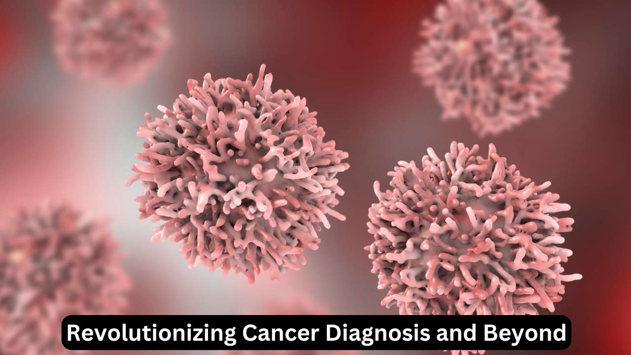
The AI model’s ability to identify diseases in tissue samples in minutes opens new avenues in cancer diagnosis and other genetic disorders. For instance, biopsy images that typically take hours to interpret can now be analyzed almost instantaneously, helping to deliver faster patient outcomes.
Enhancing Cancer Detection
Cancer diagnosis often requires early detection to improve survival rates. This AI model enables rapid screening of tissue samples for signs of cancerous cells, potentially catching cancer in its earliest stages. By processing images from diverse cancer studies, including breast and prostate cancer, the model has demonstrated proficiency in detecting tumor markers with remarkable speed and accuracy.
Applications in Veterinary Medicine
Apart from human pathology, this AI tool is also showing promise in veterinary applications. WSU is actively collaborating with veterinary researchers to utilize the model in diagnosing disease in deer and elk tissues, paving the way for broader use in animal health. By expanding AI applications beyond human medicine, researchers are helping to ensure healthier animal populations, which in turn, can improve ecosystem health and agricultural outcomes.
The Technology Behind WSU’s AI Model: Deep Learning in Action
Neural Networks and Backpropagation
The WSU AI model leverages neural networks, complex systems inspired by the structure of the human brain. When the AI misidentifies a tissue pattern, it “learns” from the error and adjusts its network using a method called backpropagation, fine-tuning its detection accuracy. This continuous learning mechanism makes the AI model increasingly precise over time.
Managing Large-Scale Data in Pathology
Handling high-resolution images and large datasets has traditionally been a challenge in pathology, with typical computers struggling to manage the size and complexity of gigapixel images. However, WSU’s AI model is designed to break down these images into manageable sections while retaining the contextual details, making it efficient in processing even the largest data inputs.
Advantages of AI in Diagnosing Tissue Pathology Over Human Pathologists
AI’s edge in tissue pathology is not merely speed; it’s about consistent accuracy, reliability, and the ability to manage high-volume data effectively. Here are some key advantages of AI in diagnosing tissue pathology:
- Reduced Time for Diagnosis: The AI model processes images within minutes, as opposed to the hours or days required by human experts.
- Decreased Human Error: While human fatigue can lead to diagnostic inconsistencies, AI provides a uniform level of performance.
- Enhanced Precision: By analyzing tissue at the molecular level, AI identifies subtle changes that are often imperceptible to the human eye.
- Scalability for Large Studies: The AI system is designed to handle vast datasets, which is essential for large-scale research projects.
Addressing Challenges and Solutions in AI-Driven Pathology
Problem 1: High Data Processing Requirements
Handling high-resolution images in gigapixel format requires immense computing power, posing challenges in data processing. To address this, the WSU team optimized their AI model to process images in smaller tiles, reducing computational strain while maintaining detail accuracy.
Problem 2: Need for Comprehensive Training Data
An AI model’s performance depends on the quality and diversity of its training data. The WSU AI model was trained on images from past epigenetic studies and cancer datasets, giving it a robust foundation to accurately diagnose multiple tissue types and diseases. Continual updating of the model with new data ensures its diagnostic capabilities remain relevant.
Problem 3: Limited Awareness Among Medical Professionals
Despite its potential, AI adoption in pathology faces resistance due to a lack of awareness or skepticism among healthcare providers. Educating medical staff on the efficacy and reliability of AI in diagnosing tissue pathology is essential to encourage its adoption.
Frequently Asked Questions (FAQs)
Q1: How does AI improve the speed of diagnosing tissue pathology?
A: AI processes tissue images at a faster rate than humans, reducing the time required for diagnosis from months to mere weeks.
Q2: What are gigapixel images, and why are they important in tissue pathology?
A: Gigapixel images are high-resolution images with billions of pixels, enabling detailed examination of tissue at a microscopic level.
Q3: How accurate is AI in comparison to human pathologists?
A: Studies show that AI can be more accurate than human pathologists, often identifying abnormalities that may be missed by human experts.
Q4: Can AI detect all types of diseases in tissue samples?
A: While AI can detect many diseases, its accuracy depends on the training data. It’s especially effective in detecting cancer and genetic disorders.
Q5: What are the applications of this AI technology in veterinary medicine?
A: WSU’s AI model is being used to diagnose diseases in animal tissue, with applications in both veterinary care and wildlife health.
Conclusion: The Future of AI in Diagnosing Tissue Pathology
The advancement of AI in diagnosing tissue pathology is a remarkable leap forward in both medical and scientific research. The deep learning model developed by Washington State University exemplifies how AI can revolutionize diagnostics by delivering quicker and more accurate results than traditional methods. With its capacity to analyze gigapixel images, detect subtle molecular changes, and improve the efficiency of large-scale studies, AI in diagnosing tissue pathology holds significant promise for the future of healthcare. Please follow out blog Technoloyorbit.

